


Patellar Dislocation/Patellofemoral Dislocation
The patella (kneecap) is a small bone that lies over and forms a part of the knee joint. attached to the quadriceps muscles of the thigh by quadriceps tendon. It attaches with the femur bone to form a patellofemoral joint. The patella is attached to the quadriceps muscles of the thigh by quadriceps tendon and protected by the medial patellofemoral ligament (MPFL) that secures the kneecap, preventing it from gliding out of the knee joint.
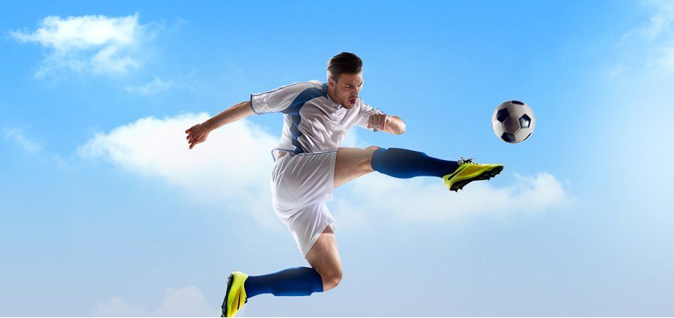
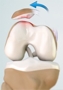
Dislocation of the patella occurs when the patella moves out of the patellofemoral groove (called as trochlea) onto the bony head of the femur. If the kneecap partially comes out of the groove, it is called subluxation, and if the kneecap completely comes out, it is called dislocation (luxation). Patella dislocation is commonly observed in young athletes between 15 and 20 years, commonly affecting women because of the wider pelvis that creates a lateral pull on the patella.
Some of the causes for patellar dislocation include a direct blow or trauma, twisting of the knee while changing direction, muscle contraction and congenital defects. It also occurs when the MPFL tears. The common symptoms include pain, tenderness, swelling around the knee joint, restricted movement of the knee, numbness below the knee and discoloration of the area where the injury has occurred.
Your doctor will examine your knee and suggest diagnostic tests such as X-ray, CT scan and MRI scan.
There are nonsurgical and surgical ways of treating patellofemoral dislocation.
The nonsurgical or conservative treatments include:
- PRICE (protection, rest, ice, compression and elevation)
- Nonsteroidal anti-inflammatory drugs and analgesics, to treat pain and swelling
- Braces or casts to immobilise the knee and allow the MPF ligament to heal
- Appropriate footwear, to control gait while walking or running, and reduce pressure on the kneecap
- Physiotherapy to control pain and swelling, prevent formation of scar of soft tissue and help in collagen formation. A physiotherapist will extend your knee and apply direct lateral to medial pressure to the knee to help in relocation. Strengthening exercises of the hip muscles and other exercises are taught to improve range-of-motion.
Surgical treatment is recommended for recurrent patella dislocation. Some of the surgical options include:
Lateral-release
It is done to loosen or release the tight lateral ligaments that pull the kneecap from its groove which increases pressure on the cartilage and causes dislocation. In this procedure, the ligaments that tightly hold the kneecap are cut using an arthroscope.
Medial patellofemoral ligament reconstruction
In this procedure, the torn MPF ligament is removed and reconstructed using grafts. Grafts are usually harvested from the hamstring tendons, located at the back of the knee and are fixed to the patella tendon using screws. The grafts are either taken from the same individuals (autograft) or from a donor (allograft). This procedure is also performed using an arthroscope.
Tibia tubercle realignment or transfer
Tibia tubercle is a bony attachment below the patella tendon which sits on the tibia. In this procedure, the tibia tubercle is moved towards the centre and then held by two screws. The screws hold the bone in place and allow faster healing, preventing the patella to slide out of the groove. This procedure is also performed using an arthroscope.
After the surgery, your doctor will suggest the use crutches for a few weeks, prescribe medications to control pain and swelling, and recommend physiotherapy, which will help you to return to your sports activities at the earliest.