


Shinbone Fractures
The tibia or shinbone is a major bone of the leg that connects the knee to the ankle. A tibial fracture is a break in the continuity of the shinbone.
There are two types of shinbone fractures
- Fractures of proximal tibia: A proximal tibial fracture is a break in the upper part of the shinbone or tibia. They may or may not involve the knee joint. Fractures that enter the knee joint may cause joint imperfections, irregular joint surfaces, and improper alignment in the legs. This can lead to joint instability, arthritis and loss of motion. These fractures are caused by stress or trauma or in a bone already compromised by disease, such as cancer or infection. Proximal tibial fractures can result in injury to the surrounding soft tissues including the skin, muscles, nerves, blood vessels and ligaments.
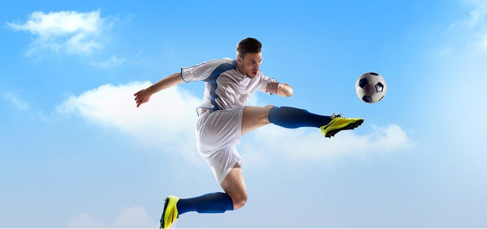
- Tibial shaft fractures: A tibial shaft fracture is a break that occurs along the length of the tibia or shinbone. These fractures can occur while playing sports such as soccer and skiing.
The symptoms of a tibial fracture include painful weight-bearing movements, tenseness around the knee, limitation of movement and deformity around the knee. Sometimes, you may have impaired blood supply secondary to the fracture, which may result in pale or cool feet. You may also experience numbness or a feeling of ‘pins and needles’ in the foot as a result of associated nerve injury.
The diagnosis of tibial fracture is based on your medical history including the history of any previous injuries, complete physical examination and imaging studies. Your doctor will evaluate the soft tissue around your joint to identify any signs of nerve or blood vessel injury. Multiple X-rays and other imaging studies, such as CT and MRI scans, may be ordered to identify the location and severity of the fracture.
The management of the fracture is based on the severity of the fracture, your medical condition and lifestyle.
Nonsurgical treatment comprises of immobilising the fractured site with the help of casts or braces to prevent weight-bearing and help in the healing process. X-rays are taken at regular intervals to assess the healing process. Weight-bearing and movement are initiated gradually, depending on the nature of the injury and your condition.
Surgical treatment is considered, to maintain the alignment of the fractured bone. External or internal fixators may be used to align the fractured bone segments. If the fracture does not involve the knee joint, rods and plates can be used. If the fracture involves the knee joint, a bone graft may be implanted to prevent the knee joint from collapsing. An external fixator is fixed when the surrounding soft tissue is severely damaged.
As the tibial fracture usually involves a weight-bearing joint, it may cause long-term problems such as loss of knee motion, instability, or long-term arthritis. Hence, a rehabilitation program is initiated along with the treatment, where you will be instructed on weight-bearing, knee movements and the use of external devices such as braces.