


Transient Osteoporosis of the Hip
Transient osteoporosis of the hip is a rare condition that causes bone loss temporarily in the upper part of the thighbone (femur). It is mostly found in young or middle-aged men between the ages of 30 and 60, and women in their later stages of pregnancy or early postpartum period (following childbirth). It is characterised by the abrupt onset of pain that increases with activity.
The hip joint is a ball-and-socket joint. A part of the pelvis bone known as the acetabulum forms the socket and the upper end of the femur, known as the femoral head, forms the ball. In patients with transient osteoporosis of the hip, the femoral head loses its density and strength, and becomes more prone to breaking.
Causes
An exact cause is unknown. Some of the proposed causes include atypical mechanical stresses acting on the hip joint, hormonal abnormalities and blockage of some of the small blood vessels surrounding the hip joint.
Symptoms
Symptoms may include:
- Unknown pain in the hip not triggered by any previous accident or injury
- Abrupt onset of pain in the anterior thigh, the side of the hip, groin or buttocks
- Pain that increases with activities or weightbearing and decreases with rest
- Intense pain with extreme hip range-of-motion
- Gradually increasing pain that becomes disabling over a few weeks or months
- A prominent limp
- Bone marrow oedema (fluid build-up in the bone marrow causing inflammation)
Diagnosis
The diagnosis of transient osteoporosis of the hip often begins with a history and physical examination. Your doctor may ask you questions related to your general health and any previous accidents or injuries. You will be asked to perform various range-of-motion exercises to replicate your pain. You may experience acute pain with weight bearing and active range-of-motion, and minimal pain when your doctor moves your hip for you (passive range-of-motion). This is one of the indicators in the diagnosis of transient osteoporosis of the hip.
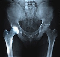
You will also be recommended to undergo imaging studies such as X-rays, CT scans, MRIs, or nuclear scans to further document transient osteoporosis of your hip.
MRI scans have been found to be particularly beneficial in documenting bone marrow oedema and are one of the most practicable studies in the diagnosis of transient osteoporosis of the hip.
Treatment
Transient osteoporosis of the hip resolves on its own and treatment involves preventing any damage to the weakened bones and minimising the symptoms and discomfort. Treatment includes:
- Medication: Nonsteroidal anti-inflammatory medications (NSAIDs) may be recommended to alleviate inflammation and pain
- Restricted weightbearing: You may be recommended to restrict or to completely avoid putting weight on your hip joint. You may need to use walking aids such as crutches, cane or a walker to limit the stress on your hip bone.
- Physiotherapy: Your doctor may instruct you on special exercises to help strengthen the muscles supporting your hip. Water exercises have been found to be helpful as they ease movement and relieve weightbearing.
- Nutrition: Vitamin D and calcium have been found to be effective in healing and rebuilding bones. Your doctor will recommend foods or supplements that can help you recover faster.